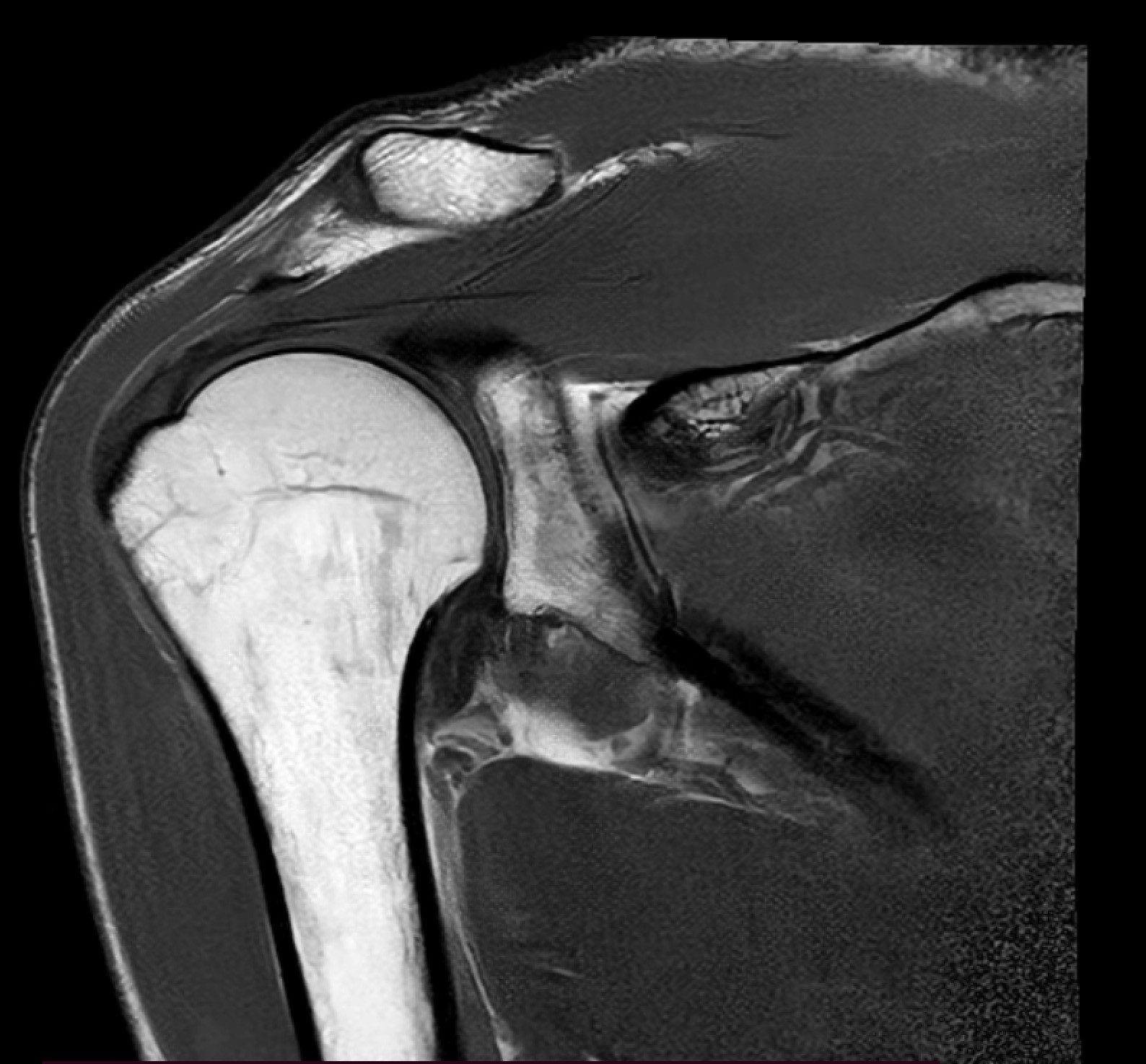
Shoulder MRI — rotator cuff tear
A rotator cuff tear is one of the most common shoulder conditions in adults. MRI is the gold standard for diagnosis — it shows tendon damage with millimetre precision.
Anatomy of the rotator cuff
The rotator cuff consists of four muscles and their tendons:
- The supraspinatus muscle — the most commonly injured (approx. 70% of cases)
- Infraspinatus muscle — external rotation
- Teres minor muscle — external rotation
- Subscapularis muscle — internal rotation
Classification of injuries
| Type | Characteristics | Treatment |
|---|---|---|
| Tendinopathy | Inflammation of the tendon | Conservative |
| Partial tear | PASTA (partial-thickness articular surface tendon avulsion) | Conservative or arthroscopy |
| Full-thickness | Small (<1cm), medium (1–3cm), large (3–5cm), massive (>5cm) | Arthroscopic repair |
| Retraction | Withdrawal of the tendon from the insertion site | Difficult repair, may require a graft |
Indications for shoulder MRI
- Shoulder pain lasting >6 weeks despite conservative treatment
- Limited range of motion
- Clicking/cracking during movement
- Weakness (Jobe’s test, Hawkins’ test)
- Following injury (fall, strain)
- Athletes (tennis, basketball, swimming, hockey)
- Before planned surgery
What an MRI of the shoulder shows
The standard APERTA protocol for the shoulder includes:
- T1 sagittal — anatomy, muscle atrophy
- T2 fat-saturated oblique coronal — rotator cuff lesions (gold standard)
- PD fat-saturated axial — cartilage, labrum
- T2 sagittal — subacromial space
MR arthrography of the shoulder — when required
MRI arthrography (MRA) with gadolinium injection into the joint is performed in cases of:
- Suspected labral tear (SLAP, Bankart)
- Shoulder instability
- Small partial rotator cuff tears not visible on standard MRI
- Overhead athletes
After an MRI — what next?
The shoulder MRI results will be interpreted by:
- Orthopaedic surgeon — decision on conservative vs surgical treatment
- Physiotherapist — a rehabilitation programme tailored to the injury
- Orthopaedic surgeon — if arthroscopy is indicated
Shoulder pain?
1T open MRI with no queues. Appointment within 3–5 days, results within 24–48 hours.
Book a shoulder MRI →