MRI Examinations at APERTA — Complete Catalog
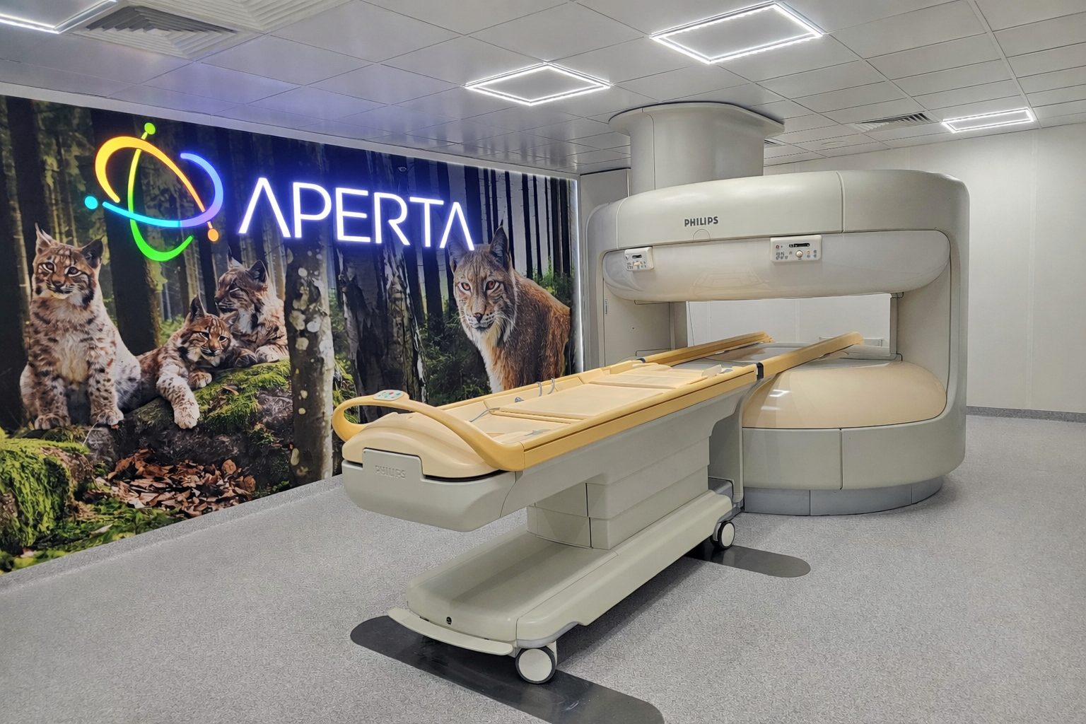
Philips Panorama 1.0 T HFO open MRI — Poland’s first high-field open MRI system. A full range of diagnostic exams for outpatients and inpatients. All protocols are available without contrast and with gadolinium contrast (if clinically indicated).
🧠 Brain and Head
Routine brain MRI
Basic examination including T1, T2, FLAIR, and DWI sequences in three planes. The first choice for headaches, dizziness, and neurological disorders.
Indications:- Headaches of unknown cause
- Suspected stroke or hemorrhage
- Epilepsy, seizures
- Multiple sclerosis (MS)
- Dementia, Alzheimer’s disease
Duration: 20–30 min · Contrast: optional (if inflammatory or neoplastic changes are suspected)
Brain MRI with gadolinium contrast
Standard examination + post-contrast T1 sequences with fat saturation. Essential when neoplasms, infections, MS, or post-surgical changes are suspected.
Indications:- Brain tumors (primary, metastatic)
- Multiple sclerosis – assessment of activity
- Inflammatory processes, abscesses
- Hemangiomas, vascular malformations
- Postoperative follow-up
Duration: 35–45 min · Contrast: intravenous (gadolinium)
MR angiography of the cerebral arteries
A non-contrast TOF (Time-of-Flight) sequence shows the circle of Willis, the internal carotid arteries, and the anterior, middle, and posterior cerebral arteries.
Indications:- Intracranial aneurysms
- Arterial stenosis, atherosclerosis
- After a TIA or stroke
- Headaches with aura
Duration: 15–20 min · Contrast: no contrast (TOF)
Cerebral MR phlebography
Imaging of cerebral venous sinuses using the venous TOF method. Detection of sinus thrombosis and venous flow dynamics.
Indications:- Suspected sinus thrombosis
- Headaches with optic disc edema
- Idiopathic intracranial hypertension
Duration: 20 min · Contrast: optional
Sella turcica (pituitary) MRI
Thin-slice T1 and T2 sequences dedicated to the pituitary region. With dynamic contrast for microadenomas.
Indications:- Hyperprolactinemia
- Cushing’s disease
- Acromegaly
- Hypopituitarism
Duration: 25 min · Contrast: dynamic gadolinium
MRI of the cranial nerves (VII, VIII)
High-resolution 3D CISS / FIESTA / DRIVE sequences showing the nerves at the cerebellopontine angle.
Indications:- Unilateral hearing loss
- Tinnitus
- Facial nerve palsy
- Acoustic neuroma (VIII)
Duration: 25 min · Contrast: gadolinium
MRI of the paranasal sinuses
Fat-saturated T1 and T2 sequences showing sinus mucosa, polyps, and abscesses.
Indications:- Chronic sinusitis
- Polyposis
- Sinus complications (orbital abscesses)
- Sinus tumors
Duration: 20 min · Contrast: optional
Orbital MRI
2–3 mm thin-slice images of the orbits in T1, T2 fat-saturated, and post-contrast sequences. Evaluation of the optic nerve and extraocular muscles.
Indications:- Optic neuritis (e.g., MS)
- Graves’ disease
- Orbital tumors
- Exophthalmos
Duration: 25 min · Contrast: gadolinium
MRI of the salivary glands
Imaging of parotid, submandibular, and sublingual salivary glands. T1, T2, DWI, post-contrast sequences.
Indications:- Salivary gland tumors (e.g., pleomorphic adenoma)
- Sjögren’s syndrome
- Salivary stones
- Inflammation
Duration: 25 min · Contrast: optional
🦴 Spine
MRI of the cervical spine (C1-C7)
Sagittal T1, T2, and axial T2 sequences at each level. Evaluation of the discs, spinal cord, and nerve roots.
Indications:- Neck pain radiating to the arm
- Numbness in the hands
- Cervical disc herniation
- Cervical myelopathy
Duration: 25 min · Contrast: optional (if spinal cord lesions, MS, or tumors are suspected)
MRI of the thoracic spine (Th1-Th12)
The entire thoracic spine in sagittal and axial sequences at clinically significant levels.
Indications:- Back pain in the thoracic region
- Scoliosis
- Osteoporotic vertebral fractures
- Metastatic lesions
Duration: 25 min · Contrast: optional
MRI of the lumbosacral spine (L1-S1)
The most commonly performed spine examination. Sagittal T1, T2, and axial T2 images at every level from L1/L2 to L5/S1.
Indications:- Lumbago, sciatica
- Lumbar disc herniation
- Spinal stenosis
- Spondylosis, spondylolisthesis
- Cauda equina syndrome
Duration: 25 min · Contrast: optional (after spinal surgery, suspected inflammation)
MRI of the entire spine
Imaging of the cervical, thoracic, and lumbar spine in a single exam. Ideal for MS, metastatic lesions, and assessment of the entire spine.
Indications:- Multiple sclerosis (MS)
- Metastatic disease
- Multisegmental scoliosis
- Post-traumatic
Duration: 50–60 min · Contrast: gadolinium-based (especially MS, oncology)
Functional spinal MRI (flexion/extension)
Examination in neutral position + flexion + extension. Shows instability, dynamic narrowing of the spinal canal.
Indications:- Cervical/lumbar spine instability
- Dynamic spondylolisthesis
- After spinal injuries
Duration: 35 min · Contrast: no contrast
🦵 Joints
MRI of the knee joint
PD fat-saturated sagittal, coronal, and axial sequences + sagittal T1. Evaluation of menisci, cruciate ligaments, and cartilage.
Indications:- Meniscus injury
- ACL/PCL/MCL/LCL tear
- Chondromalacia patella
- Joint effusion, Baker’s cyst
- Patellar tendinopathy (jumper’s knee)
Duration: 25 min · Contrast: none (MR arthrography optional)
MRI of the shoulder joint
Oblique coronal, axial, and sagittal sequences. Evaluation of the rotator cuff, labrum, ligaments, and cartilage.
Indications:- Rotator cuff injury
- Subacromial impingement syndrome
- Shoulder dislocation
- SLAP lesion (with MR arthrography)
- AC joint separation
Duration: 25 min · Contrast: none / MR arthrography with intra-articular gadolinium
Hip MRI
Imaging of the femoral head, acetabulum, labrum, and muscles. Assessment of avascular necrosis, FAI, and dysplasia.
Indications:- Aseptic necrosis of the femoral head
- FAI (femoroacetabular impingement)
- Acetabular labral tear
- Hip dysplasia
Duration: 25 min · Contrast: MR arthrography (for the labrum)
MRI of the elbow joint
3-mm thin slices in three planes. Evaluation of tendons, ligaments, cartilage, and the ulnar nerve.
Indications:- Tennis elbow (lateral epicondylitis)
- Golfer’s elbow (medial epicondylitis)
- Ulnar nerve entrapment syndrome
- Ligament injury
Duration: 25 min · Contrast: optional
MRI of the wrist
High-resolution 3D sequences for the TFCC, carpal ligaments, and cartilage. Dedicated coil.
Indications:- TFCC injury
- Carpal tunnel syndrome
- Scaphoid fracture
- Kienböck’s disease
Duration: 25 min · Contrast: none / MR arthrography
MRI of the ankle joint
PD and T2 fat-saturated sequences in all planes. Evaluation of tendons, ligaments, and the talus.
Indications:- Ankle sprains
- Osteochondral lesions (OCD)
- Achilles tendinopathy
- Fibular groove syndrome
Duration: 25 min · Contrast: none
Foot MRI
Thin 3D slices of the metatarsus, phalanges, and plantar fascia.
Indications:- Plantar fasciitis
- Metatarsal stress fracture
- Morton’s neuroma
- Freiberg's disease (necrosis of the second metatarsal)
Duration: 25 min · Contrast: optional
MRI of the hand / fingers
Small coil + high-resolution sequences. Evaluation of flexor/extensor tendons, MCP, IP.
Indications:- Rheumatoid arthritis (RA)
- Psoriatic arthritis
- Tendinopathies
- Soft tissue tumors
Duration: 30 min · Contrast: gadolinium-based (RA, inflammation)
MRI of the temporomandibular joint (TMJ)
2-mm thin slices with the jaw in the closed and open positions. Evaluation of the articular disc and ligaments.
Indications:- Jaw joint clicking
- TMJ pain
- Dislocation of the articular disc
- Bruxism
Duration: 25 min · Contrast: none
MR arthrography (intra-articular contrast)
The gold standard for the glenohumeral/hip joint labrum. Injection of gadolinium into the joint under ultrasound guidance prior to MRI.
Indications:- Shoulder labral tears (SLAP, Bankart)
- Hip acetabular labrum injury
- Wrist TFCC
- Joint instability
Duration: 45 min · Contrast: intra-articular gadolinium
💪 Muscles and soft tissues
MRI of the thigh muscles
Coronal STIR and axial T2 fat-saturated sequences of the entire thigh. Evaluation of the quadriceps, biceps femoris, and adductors.
Indications:- Muscle strains/tears (hamstring strain)
- Muscle hematoma
- Atherosclerosis
- Muscular dystrophies
Duration: 25 min · Contrast: optional
MRI of the calf muscles
Gastrocnemius muscle, soleus muscle, Achilles tendon. STIR + T1 + T2.
Indications:- Achilles tendon injury
- Tennis leg (rupture of the gastrocnemius muscle)
- Compartment syndrome
Duration: 25 min · Contrast: none
MRI of neck soft tissues
T1, T2 fat-saturated, and post-contrast sequences. Evaluation of the thyroid, lymph nodes, and laryngopharyngeal region.
Indications:- Neck tumors, lymph nodes
- Thyroid diseases
- Inflammation of the salivary glands
- Following soft tissue injuries
Duration: 30 min · Contrast: gadolinium