The diagnostic value of open 1 Tesla MRI compared to closed 1.5 T scanners
1. Introduction and clinical context
Traditional MRI scanners operating at 1.5 T and 3 T have a closed tunnel design (gantry diameter ~60 cm), which causes significant difficulties in performing the scan for some patients. It is estimated that 4–30% of patients experience significant claustrophobic discomfort during MRI, and 1–15% interrupt the scan or withdraw from it entirely1,2. The situation is particularly critical in children, where the proportion of incomplete scans without sedation may exceed 30% in patients under 7 years of age3.
Open MRI scanners, historically operating at low field strengths (0.2–0.6 T), provided comfort but their diagnostic value was limited. The current generation of open 1 T high-field systems (including the Philips Panorama 1.0T HFO) is changing this paradigm – they combine the comfort of an open design with imaging quality comparable to that of 1.5 T closed systems for most clinical indications.
2. Technical characteristics of 1T HFO vs 1.5T closed systems
| Parameter | 1T open (HFO) | 1.5T closed |
|---|---|---|
| Design | Open – two magnets (top/bottom) | Tunnel ~60 cm |
| Magnetic field | 1.0 T | 1.5 T |
| SNR ratio (vs 1.5T) | ~67% (resulting from B₀ 1T/1.5T) | baseline 100% |
| Availability of multi-element coils | Yes (multi-coil arrays) | Yes |
| Advanced sequences | DWI, MRA, MRS available | Full portfolio |
| Patient space | Open on 3 sides, no tunnel | Closed tunnel |
| Acceptable body weight | Up to ~250 kg (depending on the model) | Usually up to 130–150 kg |
| Internal diameter | No limit (open) | ~60 cm |
The lower B₀ value is partially compensated for by modern coils, optimised sequences (e.g. SENSE, mDIXON) and longer acquisition times. In clinical practice, diagnostic quality is comparable for most routine4,5 examinations.
3. Diagnostic value in selected clinical areas
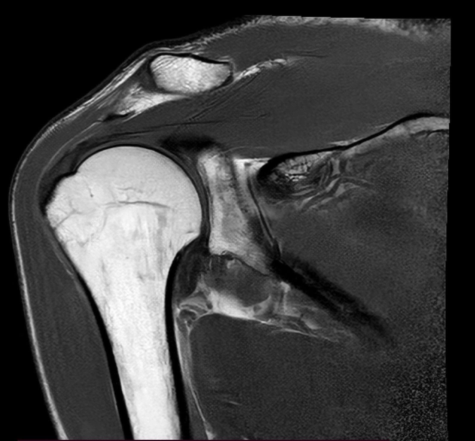
3.1 Musculoskeletal system
Comparative studies of 1T HFO versus 1.5T closed-bore MRI of the knee, shoulder and spine showed no statistically significant differences in the detection of meniscal tears, anterior cruciate ligament (ACL) tears, rotator cuff tears and disc4,6 pathologies. The sensitivity of 1T for major knee pathologies ranged from 88% to 95%, similar to that of 1.5T (90%–97%).
“High-field open MRI at 1.0 T provides diagnostic image quality comparable to 1.5 T closed-bore systems for the majority of routine musculoskeletal indications, with the added benefit of patient comfort and accessibility for claustrophobic, paediatric and bariatric populations.” (paraphrase of meta-analysis4 results)
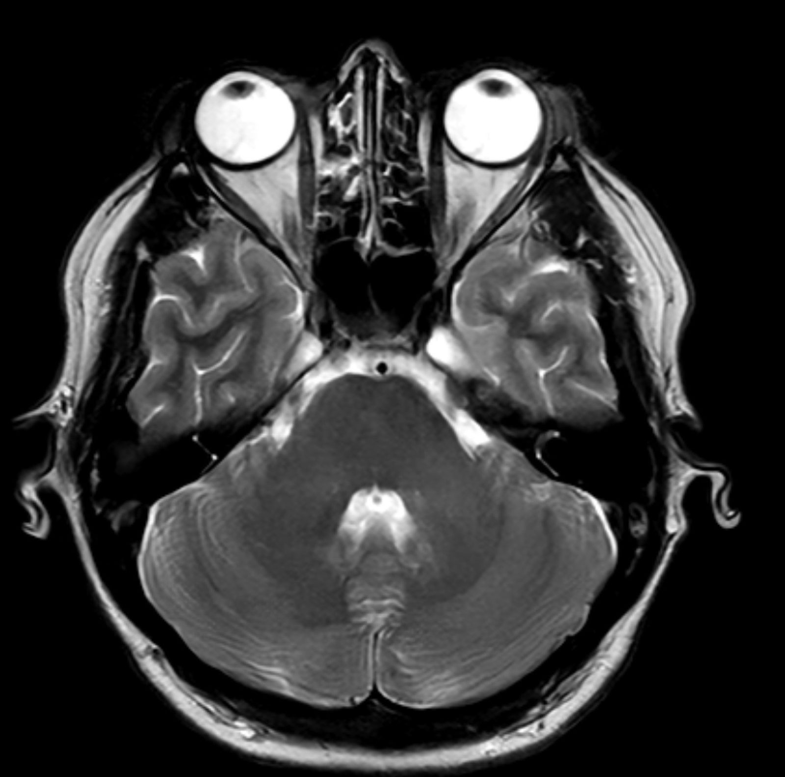
3.2 Neuroimaging
For routine brain examinations (diagnosis of strokes, epilepsy, multiple sclerosis, tumours), 1T HFO offers sufficient anatomical resolution and contrast for the vast majority of clinical7 questions. Limitations arise in highly demanding functional (fMRI), diffusion studies with high b-values (>2000 s/mm²) or high-resolution spectroscopy – in these cases, 1.5 T or 3 T should be preferred.
3.3 Paediatrics
In children, 1T HFO offers a significant clinical advantage – the presence of a parent, an open space and lower noise levels (in some models) result in a reduction in the proportion of examinations requiring general sedation. In one prospective study, the proportion of children aged 4–7 years who completed the scan without sedation increased from 38% (1.5 T closed) to 79% (open)3.
The clinical and economic implications are significant: a reduction in sedation means a reduction in anaesthetic risk, shorter hospital stays and lower costs.
3.4 Claustrophobic patients
The success rate of completing the scan in patients with claustrophobia is 96–100% in open scanners, compared with 60–85% in closed2,8 scanners. This eliminates the need for benzodiazepine premedication and the associated risks.
3.5 Larger patients / strength athletes
Closed scanners have physical limitations regarding body size (gantry ~60 cm, table weight limit 130–150 kg). Open 1T systems eliminate these limitations, enabling the diagnosis of orthopaedic patients with a bodybuilder’s physique, class II–III obesity or broad shoulders – a population for whom there is often no diagnostic alternative in MRI of comparable quality9.
4. Limitations of 1T HFO
Despite numerous advantages, one should be aware of the limitations:
- Lower SNR requires longer acquisition times for comparable resolution
- Reduced availability of advanced techniques (high-resolution DTI, 2D/3D spectroscopy, high-temporal-resolution fMRI)
- Uncertainty in the diagnosis of micropathology (e.g. cerebral microangiopathy, micrometastases <5 mm)
- Limited value in certain areas: perfusion cardiac MRI, high-resolution breast MRI – for these indications, 1.5 T or 3 T remains the preferred choice
5. Practical recommendations for referring doctors
Recommend open 1T HFO in the following cases:
- Patients with documented claustrophobia or a history of examination interruption in the tunnel
- Paediatric patients – particularly at an age where sedation would normally be required (3–7 years)
- Elderly patients with difficulty lying flat, dementia or disorientation
- Patients with an atypical body build (bodybuilders, obesity grades II–III, broad chest)
- Routine indications: musculoskeletal, neurological (excluding fMRI), abdominal MRI
Prefer 1.5 T or 3 T closed-bore in the following cases:
- Functional MRI (fMRI BOLD)
- High-resolution magnetic resonance spectroscopy
- Micropathology diagnosis (microangiopathy, micrometastases <5 mm)
- Cardiac MRI with high-resolution perfusion
- Breast MRI using the BI-RADS protocol
6. Collaboration with APERTA
The APERTA Centre (Rzeszów) offers Poland’s first high-field 1T open MRI (Philips). We collaborate with referring doctors from across the country, offering:
- Prompt appointments (24–48 hours for patients from your practice)
- A full medical report from a specialist radiologist (email within 24–48 hours)
- A dedicated communication channel with our team (doctor on call by telephone)
- Consultation support for unusual indications
Become a professional partner
Practice registration, access to a dedicated portal, preferential rates for referred patients
Check the details of the partnership →References
- Dewey M, Schink T, Dewey CF. Claustrophobia during magnetic resonance imaging: cohort study in over 55,000 patients. J Magn Reson Imaging. 2007;26(5):1322-1327. PubMed: 17969143
- Enders J, Zimmermann E, Rief M, et al. Reduction of claustrophobia with short-bore versus open magnetic resonance imaging: a randomised controlled trial. PLoS One. 2011;6(8):e23494. PubMed: 21887257
- Edwards AD, Arthurs OJ. Paediatric MRI under sedation: is it necessary? What is the evidence for the alternatives? Pediatr Radiol. 2011;41(11):1353-1364. PubMed: 21678113
- Magee T, Williams D, Mani N. Shoulder MR arthrography: which patient group benefits most? AJR Am J Roentgenol. 2004;183(4):969-974. PubMed: 15385289
- Hayashi N, Watanabe Y, Masumoto T, et al. Utilisation of low-field MR scanners. Magn Reson Med Sci. 2004;3(1):27-38. PubMed: 16093618
- Cotten A, Delfaut E, Demondion X, et al. MR imaging of the knee at 0.2 and 1.5 T: correlation with surgery. AJR Am J Roentgenol. 2000;174(4):1093-1097. PubMed: 10749258
- Rutt BK, Lee DH. The impact of field strength on image quality in MRI. J Magn Reson Imaging. 1996;6(1):57-62. PubMed: 8851404
- Spouse E, Gedroyc WM. MRI of the claustrophobic patient: interventionally configured magnets. Br J Radiol. 2000;73(866):146-151. PubMed: 10884728
- Uppot RN. Impact of obesity on radiology. Radiol Clin North Am. 2007;45(2):231-246. PubMed: 17502214
This page is intended for informational purposes for medical professionals and does not replace individual clinical assessment. The choice of diagnostic modality is always at the discretion of the patient’s attending physician. Current literature is available on PubMed (https://pubmed.ncbi.nlm.nih.gov).
Contact our medical team
Are you a doctor interested in collaboration or a clinical consultation? Fill in the form – we will reply within 24 hours.