MRI in oncology – its role in diagnosis
Magnetic resonance imaging plays a key role in the diagnosis, staging and monitoring of cancer patients. The absence of ionising radiation means that MRI can be repeated many times without the risk of cumulative dose – which is particularly important for patients requiring frequent check-ups. I will explain where MRI is the gold standard, where alternatives (CT, PET) are better, and how APERTA can support your cancer treatment plan.
In this article
Main oncological indications for MRI
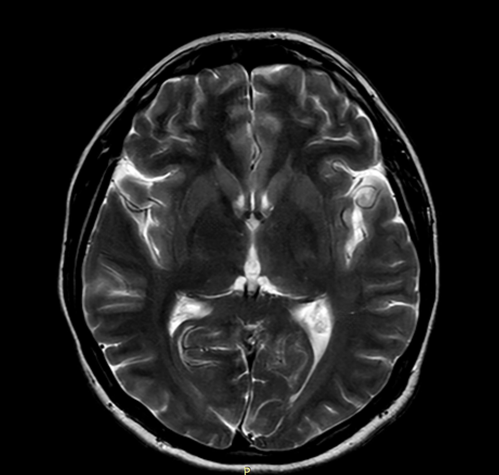
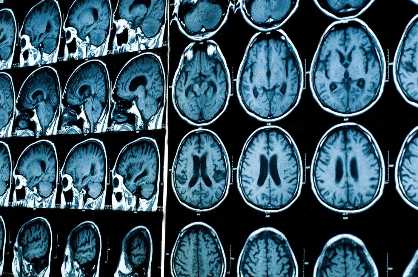
MRI is the method of choice for: tumours of the central nervous system (brain, spinal cord); primary bone and soft tissue tumours (sarcomas); tumours of the lower abdomen (prostate, endometrial, cervical and rectal cancer); breast cancer (as a supplement to mammography in high-risk cases); liver tumours (HCC, metastases); head and neck cancers. CT is preferable for: lesions of the lungs, ribs, and thoracic lymph nodes.
MRI in diagnosis vs staging vs follow-up
Diagnosis: MRI detects a tumour and assesses its nature (cystic vs solid). Staging: assesses local extent (T-staging in TNM) and involvement of adjacent structures. Follow-up: monitors response to treatment (chemo/radiotherapy) and detects early recurrences. Contrast-enhanced MRI is often superior to CT for soft tissues (better contrast between the tumour and surrounding tissue).
Contrast in oncological MRI – is it safe?
Gadolinium contrast is the standard in oncological MRI – it helps distinguish tumour tissues, assess vascularisation (markers of aggressiveness) and detect small lesions. Safety: serious allergic reactions in <0.1% of cases; nephrogenic systemic fibrosis (NSF) is very rare, mainly in patients with renal failure (eGFR <30). In patients with eGFR <30, we use MRI without contrast or CT. Gadolinium does not accumulate permanently in the body in patients with normal kidney function.
How much do repeated MRI scans cost in oncological follow-up?
Oncology patients often require an MRI every 3–6 months for years. The National Health Fund (NFZ) reimburses selected indications, but waiting times can be long (3–6 months). At APERTA, we offer oncology follow-up packages with preferential prices and a guaranteed appointment within 7 days. Contact our team for details – it is important to maintain continuity of monitoring.
APERTA and cancer patients
Our open 1T scanner is particularly valuable for oncology patients with: claustrophobia developed during treatment (chemotherapy and radiotherapy often exacerbate this); patients post-surgery who find it difficult to lie flat; elderly patients with general weakness; and palliative care patients requiring comfort. We collaborate with leading oncology centres in Poland – we can exchange DICOM images directly with your oncologist.
Frequently asked questions
Will an MRI detect all cancers?
No. MRI has its limitations – it is excellent at detecting lesions in the brain, spinal cord and pelvis, but less effective in the lungs (where CT is superior).
I’m currently undergoing chemotherapy – can I have an MRI?
Yes, an MRI does not affect chemotherapy. However, please inform us if you are currently undergoing treatment so that we can adjust the protocol.
Will an MRI detect an early tumour recurrence?
Often yes, if the protocol is correctly selected. That is why regular check-ups are crucial.
I have a port-a-cath – can I have an MRI?
Yes, most ports are MRI-compatible. Check the device documentation.
Can I stop chemotherapy for the duration of the MRI scan?
There is no need to. An MRI does not interfere with chemotherapy.
Should I be concerned about contrast after multiple scans?
In patients with normal kidney function, gadolinium accumulation does not cause any known clinical problems.
This content is for information purposes only and does not replace a medical consultation.