Brain MRI – when is it recommended?
A brain MRI is the most accurate imaging test of the central nervous system. It shows the brain’s structure with sub-millimetre resolution, detecting changes that are not visible on a CT scan. I will explain when a doctor recommends a brain MRI, which conditions it detects best, and how to interpret typical radiological findings.
In this article
Main clinical indications
Brain MRI is the gold standard for: chronic or atypical headaches (>50 years of age with new symptoms, waking from sleep, worse in the morning, with visual/neurological symptoms); epilepsy diagnosis (exclusion of tumours, malformations); multiple sclerosis (the most sensitive method – white spots, T2/FLAIR); strokes (acute and chronic, DWI for early detection); brain tumours (characterisation, localisation); dementia (differentiation of types); head injuries (axonal lacerations, haemorrhages); infections (encephalitis, abscess).
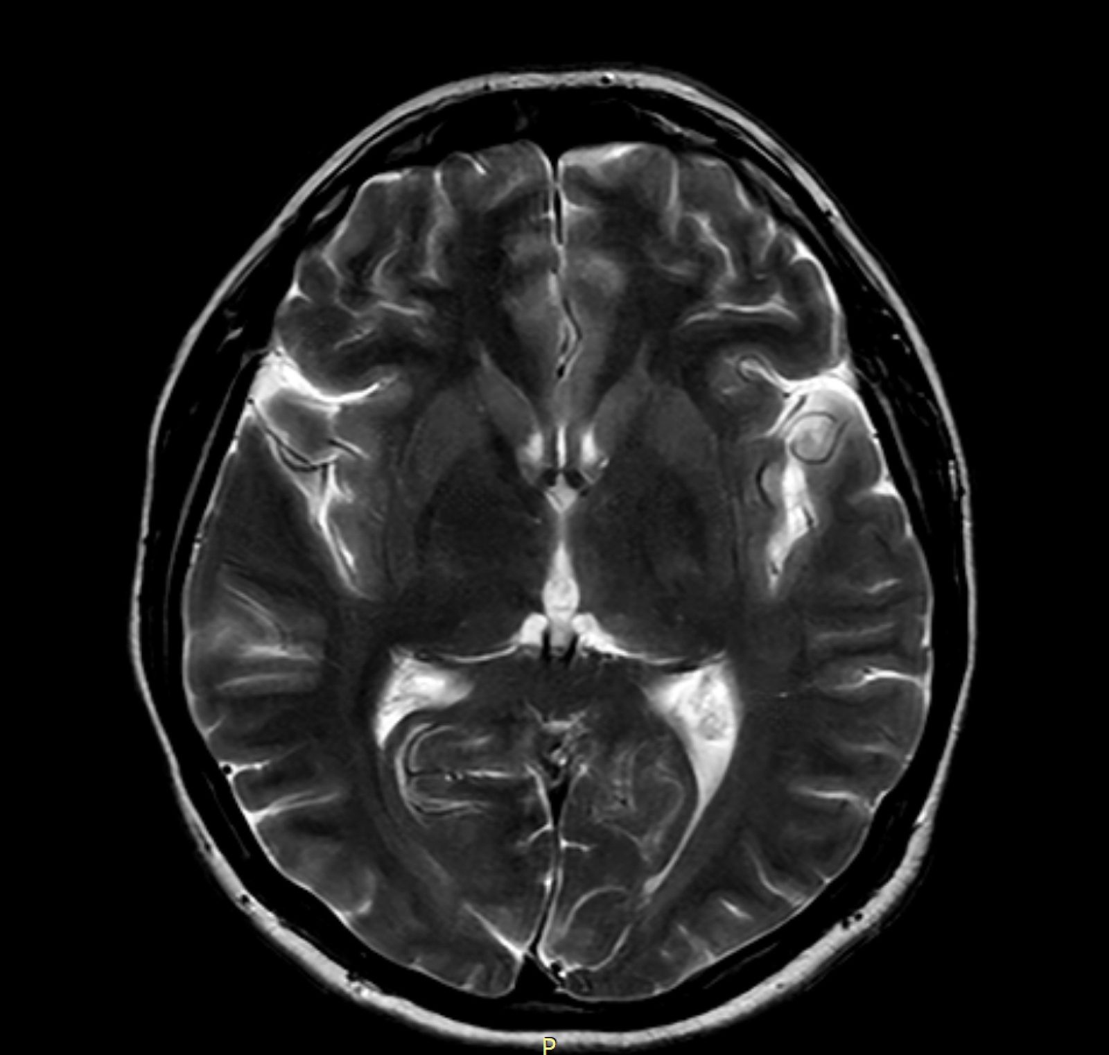
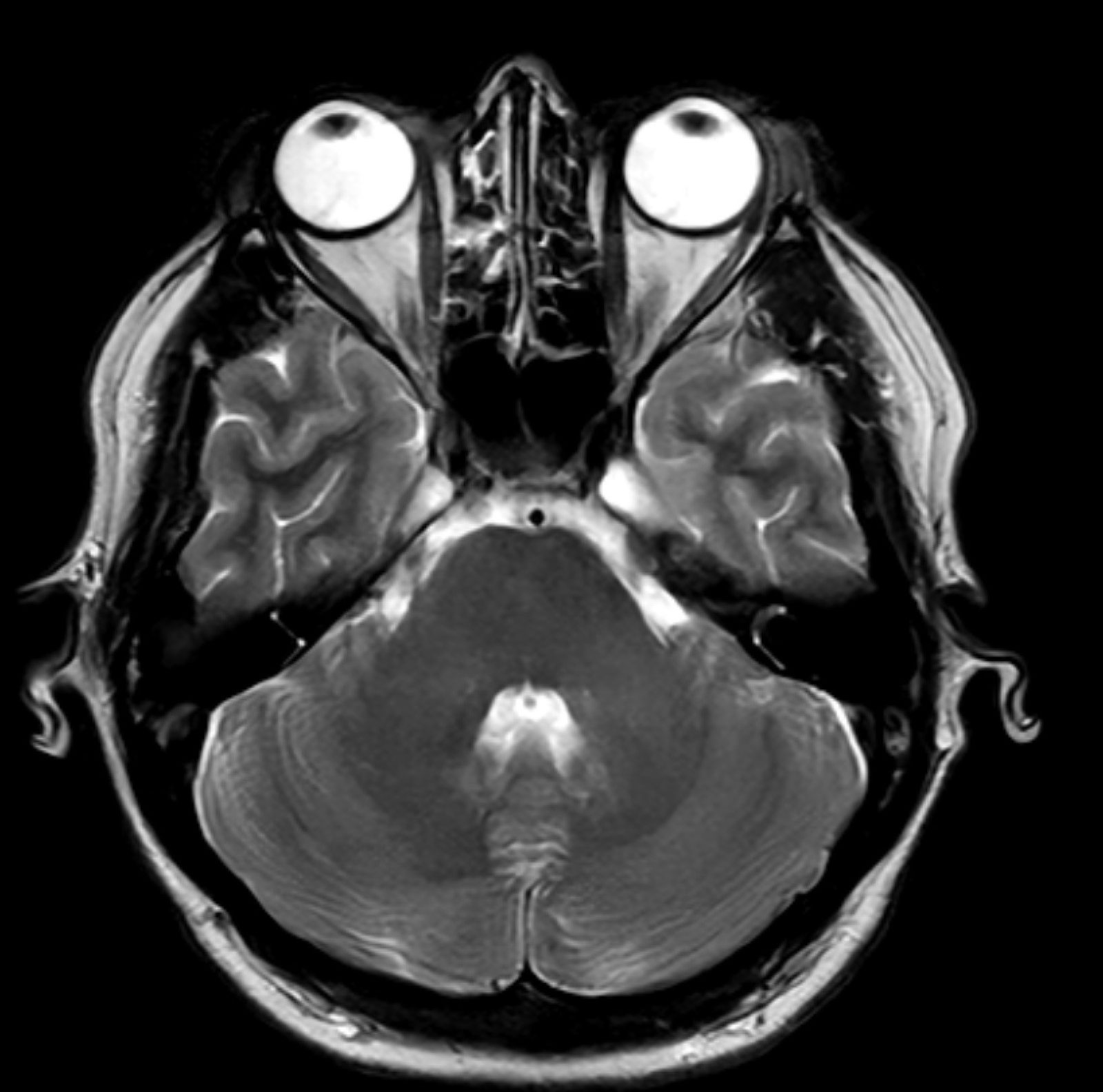
Brain MRI sequences – what they mean
T1-weighted: good anatomical definition, fat appears bright, water appears dark. With contrast (T1+Gd) = enhancement of pathology. T2-weighted: water appears bright – detects oedema, inflammatory changes, demyelination. FLAIR (Fluid Attenuated Inversion Recovery): like T2, but with CSF suppression – ideal for periventricular lesions (MS). DWI (Diffusion Weighted Imaging): detects acute ischaemic strokes within the first few hours. SWI (Susceptibility Weighted): microhaemorrhages, mineralisation. MRA (MR Angiography): vessels without contrast (TOF).
What do typical terms in the description mean
“Hyperintensity on T2/FLAIR” = bright lesion – oedema, gliosis, demyelination, MS, early stroke. “Cortical atrophy” = reduction in cerebral cortex volume – typical in ageing, dementia. “Ventricular enlargement” = hydrocephalus or tissue loss. “Vascular microangiopathy” = small lesions in the white matter in people with vascular risk factors (hypertension, diabetes). “No contrast enhancement” = usually a good sign (no active lesion). “Ring-shaped enhancement” = may indicate a tumour, abscess.
Multiple sclerosis – MRI diagnosis
MRI is key to the diagnosis of MS. McDonald criteria 2017: 1) ≥1 typical MS T2 lesion in ≥2 of 4 locations (periventricular, cortical/subcortical, subependymal, spinal cord); 2) Temporal dissemination (simultaneous enhancement + non-enhancing lesions, or a new lesion at follow-up); 3) Exclusion of alternative causes. Repeat MRI scans every 6–12 months are standard for monitoring disease activity and response to disease-modifying treatment.
Brain MRI at APERTA – comfort for neurological patients
Patients with headaches, anxiety disorders, dementia or a recent stroke often find the classic MRI tunnel more difficult to tolerate. The APERTA 1T open scanner is particularly valuable for: claustrophobia developing during the diagnostic process; patients with epilepsy (greater comfort = lower risk of a seizure); senior citizens with dementia (the option for a carer to be present); migrants with PTSD. Most routine neurological protocols can be performed on a 1T scanner with diagnostic quality comparable to that of a 1.5T scanner.
Frequently asked questions
Does a brain MRI require contrast?
It depends on the indication. Diagnosis of a tumour, active MS, infection – yes. Migraine, primary epilepsy – usually not.
How long does a brain MRI take?
The standard protocol takes 20–30 minutes; with contrast, 35–45 minutes.
Will an MRI detect all types of headache?
It detects structural causes. Primary migraine and tension headaches do not show up on an MRI.
I have tattoos and piercings on my head – can I have a brain MRI?
Tattoos are fine, but piercings must be removed.
Does an MRI scan after a stroke help predict a recurrence?
Indirectly – it shows the extent of the damage. The rehabilitation plan is drawn up by a neurologist based on clinical findings and the MRI.
Can I have an MRI scan after an epileptic seizure?
Yes, even within the first few hours – it helps rule out a tumour or haemorrhage.
This content is for information purposes only and does not replace a medical consultation.